Liver cancer has remained one of the leading causes of cancer-related mortality over the years. Furthermore, liver cancer is trending towards affecting younger populations and has very unpredictable progression. The application of AI in early diagnosis and classification of liver cancer promises to usher in a completely new era in healthcare.
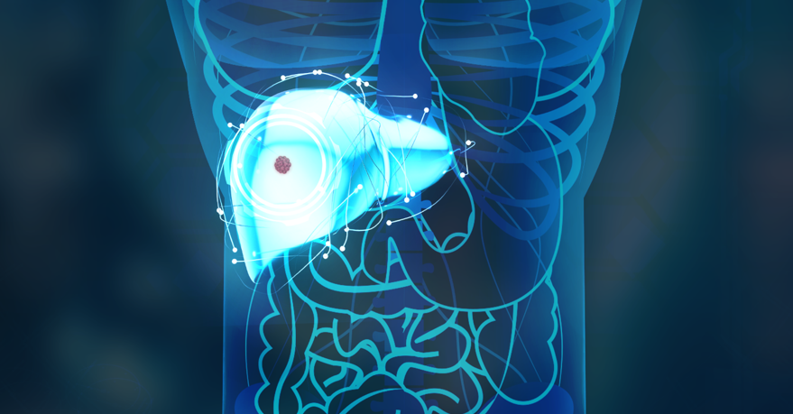
The liver is the largest internal organ in the body. It plays a crucial role in maintaining health and bodily functions by filtering out toxins, participating in digestion, storing glucose (a simple form of sugar), and releasing it into the blood when the body needs energy.
Liver cancer is the rapid growth and spread of unhealthy cells within the liver. When these cells become cancerous, the liver can no longer perform its vital functions, leading to severe consequences for the body.

Based on the origin of the disease, liver cancer is classified into two types: primary liver cancer and secondary liver cancer.
Secondary liver cancer (metastatic liver cancer) occurs when cancer cells from other organs, such as the colon, rectum, stomach, pancreas, esophagus, breast, lungs, etc., metastasize to the liver and form malignant tumors there. Secondary liver cancer cannot be cured because it represents the final stage of cancer, during which liver function is almost completely lost.
Primary liver cancer originates from the cells within the liver itself. National data indicate that this is the third most common type of cancer in Vietnam, following gastric and bronchial cancers. Primary liver cancer is further divided into four main types:
Hepatocellular carcinoma (HCC) is the most common type of liver cancer, accounting for about 80% of cases. It typically develops in individuals over 50 and is more common in men. The majority of HCC cases occur in people with chronic hepatitis B or C infections or cirrhosis due to alcohol. HCC is known for its rapid progression and can be fatal within six months.
Cholangiocarcinoma forms in the small bile ducts that transport bile to the gallbladder and then to the small intestine to aid in digestion. This type accounts for about 15% of liver cancer cases and is often found in patients with primary sclerosing cholangitis, chronic bile duct stones, or parasitic infections.
When cancer forms in the bile ducts within the liver, it is called intrahepatic cholangiocarcinoma. It is known as extrahepatic cholangiocarcinoma if it develops in the bile ducts outside the liver.
Hepatoblastoma is a rare type of liver cancer that usually occurs in children under the age of four. If treated promptly with surgery or chemotherapy, the prognosis is very favorable, with a survival rate exceeding 90% if detected early.
Hepatic angiosarcoma is a rare and aggressive cancer originating from the blood vessels of the liver, typically found in individuals over 60 years old. This cancer progresses very quickly, and it is often diagnosed at an advanced stage. Common symptoms include right upper quadrant pain, hepatomegaly, splenomegaly, anemia, and ascites.
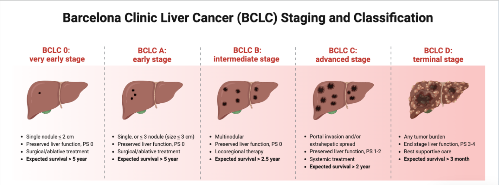
Although there are several staging systems for liver cancer worldwide, such as Okuda, CLIP, and TNM, the Barcelona Clinic Liver Cancer (BCLC) staging system is the most widely used in major treatment centers globally and in Vietnam. According to this system, liver cancer is divided into five stages based on tumor characteristics, liver function (Child-Pugh classification), and the patient's performance status (PS).

- Very Early Stage (Stage 0)
- Early Stage (Stage A)
- Intermediate Stage (Stage B)
- Advanced Stage (Stage C)
- Terminal Stage (Stage D)
Despite hepatocellular carcinoma (HCC) being the most common type of liver cancer, accounting for 80% of cases, early detection of HCC remains challenging. Current diagnostic techniques struggle to identify small lesions in the early stages, resulting in most cases being discovered at intermediate or late stages. This presents a significant barrier to early detection and treatment.
Additionally, once liver lesions are identified, traditional clinical techniques face difficulties in distinguishing between HCC and non-HCC tumors, complicating accurate treatment planning for patients.
Beyond imaging diagnostics, liver biopsy is a top choice for effectively diagnosing and treating liver tumors. However, this invasive procedure poses barriers for patients, often causing reluctance to undergo the procedure.
The global medical community has seen numerous advancements, particularly in applying artificial intelligence (AI) to imaging diagnostics. However, few institutions have successfully implemented AI solutions for liver cancer diagnosis and treatment.
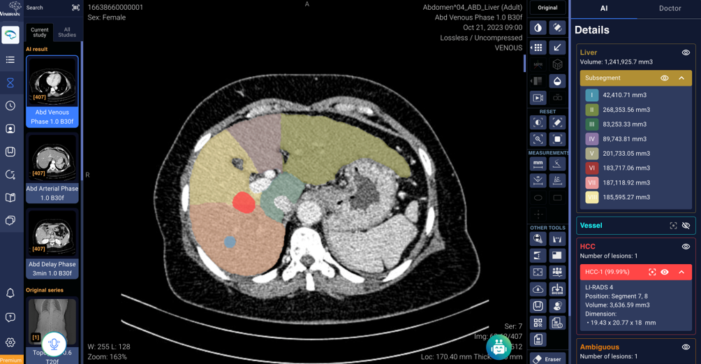
In Vietnam, the DrAid™ CT Liver Cancer platform by VinBrain has made a significant impact among medical professionals. It stands out as one of the pioneering global platforms for utilizing AI for early liver cancer detection. DrAid™ CT Liver Cancer is designed to assist radiologists by automatically detecting abnormal liver tumors as small as 5mm through CT imaging. This platform provides clinical solutions for early liver cancer diagnosis using AI and aids oncologists in developing more effective treatment plans.

Using advanced Multifaceted CT scan technology, DrAid™ CT Liver Cancer enables imaging from multiple angles and depths, detecting small lesions as tiny as 5mm that the naked eye might miss. It includes four main features:
VinBrain has achieved a significant milestone in early liver cancer detection research. Their study on a new method for processing multifaceted CT scans to improve the screening of hepatocellular carcinoma (HCC) has been published in the prestigious scientific journal Nature Scientific Reports. Scientific Reports is a leading global journal in natural sciences, psychology, medicine, and engineering. With an H-index of 282, it is the fifth most cited journal worldwide.
The breakthrough in this research lies in using radiomics features in the wavelet domain. By enhancing the visualization of liver lesions, VinBrain researchers have pioneered a method to distinguish between HCC and non-HCC tumors, significantly contributing to early detection and improved patient treatment outcomes.
Sources:
https://www.ncbi.nlm.nih.gov/pmc/articles/PMC10251861/
https://pubmed.ncbi.nlm.nih.gov/36813095/
https://benhvienungbuouhungviet.vn/kien-thuc-ung-thu-gan/chan-doan-ung-thu-gan-phan-giai-doan.html
Top