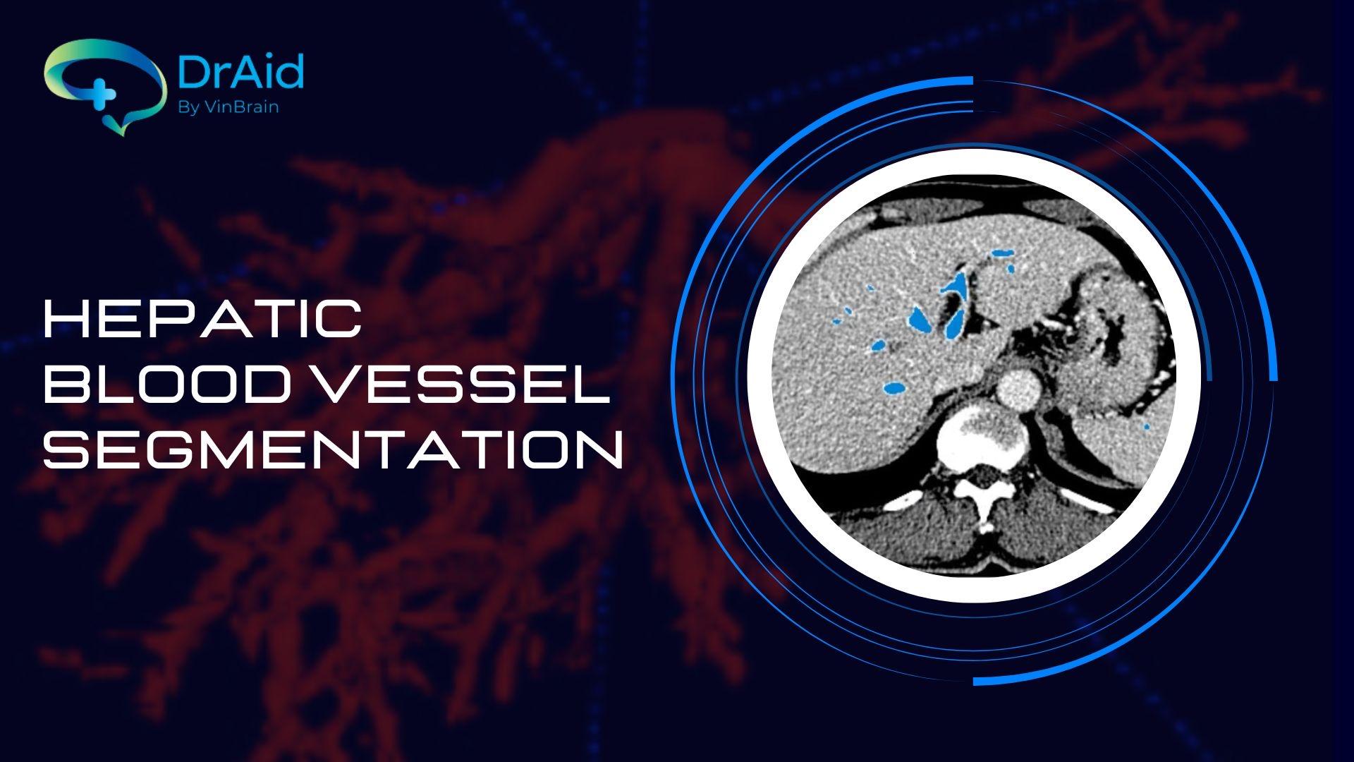
DrAid™ CT Liver Cancer Diagnosis and Treatment introduces a new feature utilizing the most cutting-edge algorithms to automatically segment liver blood vessels, which holds significant importance in the diagnosis and specialized work of oncologists when interpreting abdominal CT images of liver patients.
The liver is one of the most complex vascular structures in the body, receiving a dual blood supply from the hepatic vein and artery. The hepatic venous system includes hepatic veins and the portal vein, forming a highly intricate tree-like structure. According to anatomy, blood from the vein flows into the liver through the portal vein, then drains into the inferior vena cava through the central hepatic veins. These vessels also deliver nutrients and oxygen to nourish liver tumors.
As blood flow through the portal vein increases, blood flow through the hepatic artery decreases, and vice versa (a buffering response of the hepatic artery). This compensatory dual blood supply mechanism partly safeguards the liver against localized ischemia in healthy livers.
The portal vein is formed by the splenic vein and superior mesenteric vein. After their confluence, the portal vein directs towards the liver, branching out to the porta hepatis, where it further divides into the right and left portal veins, then entering the live into smaller branches supplying the lobes, lobules, and smaller structures.
The hepatic artery originates from the celiac trunk's visceral branch, enters the liver, and branches similarly to the portal vein. Additionally, the hepatic artery is the primary blood supply for the gallbladder.
The hepatic veins transport blood from the liver to the inferior vena cava, returning blood to the heart. There are three main hepatic veins: the right hepatic vein, middle hepatic vein, and left hepatic vein.
Specific vascular damage can occur in the hepatic artery, veins, or portal vein. Hence, hepatic vasculature segmentation is a topic of great interest in medical image analysis. Prior to treatment, physicians need a firm understanding of the liver's vascular structure. This is a premise for doctors to formulate diagnoses, and weigh treatment and surgery options, as well as evaluate clinical results for patients with hepatocellular carcinoma (HCC), a highly dangerous form of liver cancer, and patients with diverse liver lesions and tumors.
DrAid™ CT Liver Cancer Diagnosis and Treatment's new feature will assist doctors in rapidly segmenting liver blood vessels automatically, visually analyzing the precise location of tumors within the liver's vascular system, including the relationship between blood vessels and different regions of the liver, encompassing tumors. This information is vital for several treatment methods such as surgical resection and transarterial chemoembolization (TACE), as well as post-treatment evaluations.


Top


